Can not see Light/Endogeneous peptides

support
Can not see Light/Endogeneous peptides VM26  2024-03-20 18:28
 

Hi All,

I have been trying to develop a PRM assay for 1 protein for which I have 3 Heavy peptides. I have been able to develop a linear curve for LOD and LOQ using CSF (fixed amount) as a matrix with varying conc of Heavy peptides.
For Ratio, I used light as the internal standard...is that correct?

Using Light as the internal standard and heavy as Isotopically labeled, I could plot a linear graph for 1 peptide. However, I was unable to get a linear curve for 2 peptides of the same protein.
I can see the reason why as the endogenous peptides (Light peptides) might be way too low as compared to the Heavy peptide amount spiked that might be way too high for which the Ratio H/L becomes infinity.
To overcome this, I used a ratio CSF: Heavy peptide (in fmoles/ul) as 1:1.

However, I am unsure how to proceed with 1:1.
My concern is with body fluid in my case its CSF. I tried multiple way to do 1:1 ratio

CASE 1
First, i prepared different dilution of Heavy peptide standards from stock of 1mM such as 2500, 1000, 500,........0.25 fmol/ul.
I then took 5 ul from each standard and added to 5 ul of CSF and injected 1ul to column. My worry is the volume ratio 1:1 is right or not.

CASE 2
Secondly, I tried to prepare a 2500 fmol/ul from the heavy peptide stock. Took 5ul from 2500fmol/ul and added to 5 ul CSF. I prepared the dilution using this CSF+2500 Dilution as stock and diluted it down to 1000.......0.25fmol/ul. Here for every dilution, both CSF and Standard are equally getting diluted.

In Case 1; my every dilution vial has 5ul CSF (fixed volume so fixed amount of CSF) and 5ul from respective dilution tube (1000.....0.25fmol/ul)

In Case 2; my main mixed stock of 5ul CSF+ 5ul 2500fmol/ul is serving as highest stock and I am diluting using this as the main vial and 0.1% Formic acid as diluent.

Now, I am unsure which will work best as my endogenous peptides are way low but get overexpressed in the disease model of my interest.

CSF used here is the pool of 20 patients to avoid heterogeneity.

Can you suggest some solution.

Thank you.
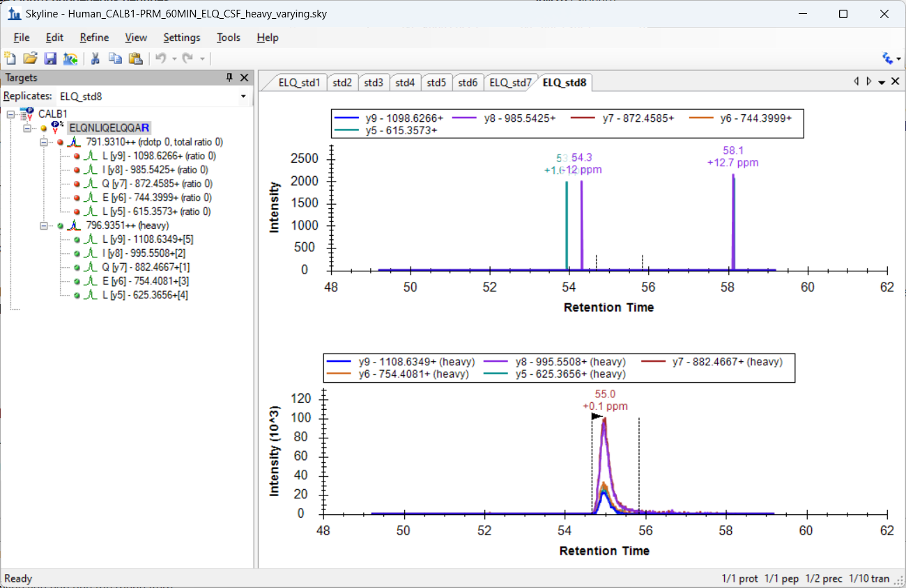
Varsha


Nick Shulman responded:  2024-03-20 19:29
I am not sure that I understand your question but if you send us your Skyline document it might make more sense.

In Skyline you can use the menu item:
File > Share
to create a .zip file containing your Skyline document and supporting files including extracted chromatograms.
If that .zip file is less than 50MB you can attach it to this support request.
You can upload larger files here:
https://skyline.ms/files.url
--Nick
 
vmohanty responded:  2024-03-21 05:22
Hi Nick,

I am attaching the file for one peptide which gave nice results and also the other 2 peptides for which i did not get any results.

I am also experiencing different RT for different transitions for peptide 2. I am not sure why. Ideal RT must be between 49-59 min. However, I am seeing different RT for different y-ion series of endogeneous light peptides.

My main idea here is to plot a Reverse calibration curve to know the LOD and LOQ of my heavy peptide to be soiked in my CSF sample. For generating the curve, I have used a pool of 20 individuals CSF and spiked different concentrations of heavy peptide.


Varsha
 
Nick Shulman responded:  2024-03-21 08:35
I can't tell what might be going wrong from looking at those Skyline documents.
If you send me some of your .raw files I might have some ideas.
Those .raw files will probably be too large to attach to this support message so you should upload them here:
https://skyline.ms/files.url
-- Nick
 
vmohanty responded:  2024-03-21 09:53
Hi Nick,

Thank you. I am uploading my raw files in the URL provided. Meanwhile, I am attaching the Instrument method and inclusion list used for my analysis.

Thank you.
Varsha
 
vmohanty responded:  2024-03-21 10:48
Hi Nick,

I have uploaded my files. Please have a look at the RAW files.
Its uploaded as Raw_Endogenous by vmohanty (Endogeneous_2024-03-20)
 
Nick Shulman responded:  2024-03-21 11:49
It looks like the concentration of the endogenous peptide is a lot lower than the heavy peptide.
You should go to "Settings > Peptide Settings > Modifications" and change "Internal Standard Type" to "None".

Skyline assumes that the Internal Standard is going to be easy to detect in all of your replicates. If the Internal Standard Type has been set to something then Skyline does all of its peak picking based on looking at the internal standard's chromatograms. In your case, the beautiful heavy chromatograms will not be used at all in terms of deciding where to set the peak boundaries which is not at all what you want.

You probably also do not want to normalize by dividing by the light areas. I would recommend going to "Settings > Peptide Settings > Quantification" and changing "Normalization method" to "None" instead of the "Ratio to Light" that you currently have it set to.
-- Nick
 
vmohanty responded:  2024-03-21 12:04
Thank you.
setting Internal standard to None and Normalization method to None, did help me.

Did you see the files I attached. I am not sure why for peptide 2 my transitions of 1 precursor have different RTs.
Is it mandatory to provide start time and End time for every peptide in the inclusion list?
 
Nick Shulman responded:  2024-03-21 12:52
I am not sure what you are asking.
Can you send me a screenshot of what you are looking at?
Also, it might be helpful if you did "File > Share" again to and send me a new .sky.zip so that I'm looking at the same Skyline document as you.
-- Nick
 
vmohanty responded:  2024-03-22 09:13
Hi Nick,

Please see the attached file.
My heavy peptide is in CSF pool. Heavy peptide concentrations are varied and CSF volume is kept constant.

In peptide settings;
As my endogenous peptide so low, I kept Internal standard as None and Normalization method as None as mentioned by you.

I am experiencing different RTs for different transitions for this peptide


Varsha
 
Nick Shulman responded:  2024-03-23 13:56
Varsha,

I am not sure what you expect things to look like.
It looks to me that your light peptide really is not present in your sample.
The chromatograms for some of your transitions have tiny blips of signal at different retention times. You can see this as different colored spikes in the upper pane of the graph in the attached screenshot.
When only one of your transition's chromatogram has signal at a particular retention time, that is an indication that the signal is coming from something other than your peptide of interest.
If the signal was coming from your peptide then each transition's chromatogram would have a peak at the same retention time.

-- Nick